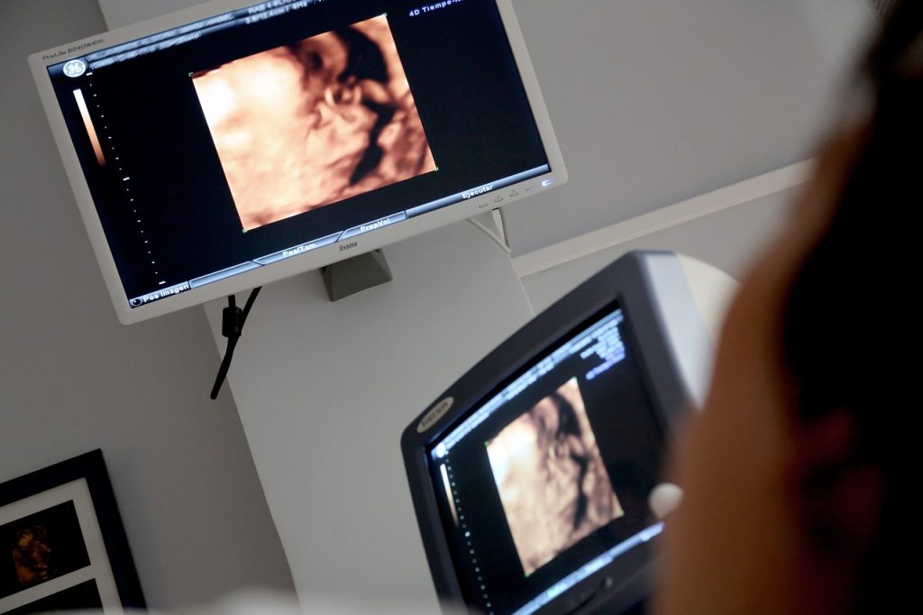
Fetal growth scan, also known as a growth ultrasound, is a medical procedure used to assess the growth and development of a fetus during pregnancy. This non-invasive procedure is typically performed between the 18th and 22nd weeks of gestation, although it can be done later in pregnancy as well. In this article, we will discuss what fetal growth scan is, why it is important, and what to expect during the procedure.

What is Fetal Growth Scan?
A fetal growth scan is an ultrasound scan that is used to measure the size and development of a fetus during pregnancy. The ultrasound technician will use a handheld device called a transducer that emits high-frequency sound waves to create an image of the fetus on a monitor. The image will show the size and position of the fetus, as well as the amount of amniotic fluid surrounding it.
During a fetal growth scan, the technician will measure several different aspects of the fetus, including its head circumference, abdominal circumference, and femur length. These measurements can help determine the estimated weight of the fetus, as well as its age and developmental stage. The technician will also look for any abnormalities or potential complications, such as an abnormal amount of amniotic fluid or the presence of multiple fetuses.
Why is Fetal Growth Scan Important?
Fetal growth scans are an important part of prenatal care because they can help detect potential problems with the fetus or the pregnancy. By monitoring the size and development of the fetus, healthcare providers can identify any issues early on and take appropriate steps to address them. This can help reduce the risk of complications and improve the outcome of the pregnancy.
One of the most important reasons to have a fetal growth scan is to identify fetal growth restriction (FGR), which occurs when the fetus is not growing at the expected rate. FGR can be caused by a variety of factors, such as placental insufficiency, maternal health problems, or genetic abnormalities. If FGR is detected, healthcare providers may recommend additional monitoring, dietary changes, or even early delivery in some cases.
Fetal growth scans can also be used to monitor the growth of fetuses that are at risk for complications, such as those with a family history of growth problems, gestational diabetes, or high blood pressure. In these cases, the scans can help detect potential problems early on and provide reassurance to the parents that the fetus is developing properly.
What to Expect During a Fetal Growth Scan?
A fetal growth scan is a non-invasive procedure that typically takes about 30 minutes to complete. The ultrasound technician will ask the mother to lie down on an examination table and expose her abdomen. A clear gel will be applied to the abdomen to help the transducer make contact with the skin and obtain clear images of the fetus.
The technician will move the transducer over the abdomen to obtain images of the fetus from various angles. The mother may be asked to change positions or move around to help the technician get a better view of the fetus. During the procedure, the technician will measure the fetus’s head circumference, abdominal circumference, and femur length to estimate its weight and developmental stage.
After the procedure, the technician will clean the gel from the mother’s abdomen and provide her with a printout of the images. The images will be reviewed by a healthcare provider, who will then discuss the results with the mother and provide any necessary recommendations or follow-up care.
In conclusion, fetal growth scans are an important part of prenatal care that can help detect potential problems with the fetus or the pregnancy. By monitoring the size and development of the fetus, healthcare providers can identify any issues early on and take appropriate steps to address them. If you are pregnant, talk to your healthcare provider about the benefits of having a fetal growth scan and whether it is recommended for your pregnancy.

